Coarctation of the aorta: Congenital heart disease is easy to miss

In Vietnam, diseases related to the aorta often cause complications with a high risk of death because patients are not detected in time. Coarctation of the aorta is not an exception. Therefore, in the article below, SignsSymptomsList hopes to provide all the necessary information to identify and treat this easily missed congenital heart disease.
content
- 1. Overview of coarctation of the aorta
- 2. Problems encountered with coarctation of the aorta
- 3. Acquired risk factors
- 4. Causes of coarctation of the aorta
- 5. Clinical symptoms by subject
- 6. Diagnosis of coarctation of the aorta
- 7. Coarctation of the aorta
- 8. Monitoring patients with coarctation of the aorta
- 9. Is coarctation of the aorta dangerous?
1. Overview of coarctation of the aorta

The aorta is the main and largest artery in the human body, has a rod shape, originates from the left ventricle of the heart, runs a U-circle around the upper chest and ends around the navel area. The aorta plays a major role in maintaining blood circulation during diastole after they are pushed into the aorta by the left ventricle during systole.
2. Problems encountered with coarctation of the aorta

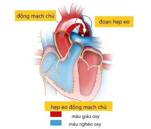
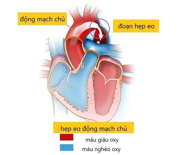
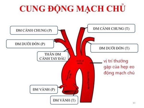
Coarctation is usually located near the origin of the left subclavian artery, which is also near the ductus arteriosus at birth.
Frequent
- Ventilation communication
- Have ductus arteriosus
- Mitral aortic valve with stenosis and regurgitation
- Turner syndrome (Statistics show that 30% of patients with Turner syndrome have coarctation of the aorta. In contrast, 5-15% of female patients with coarctation of the aorta have Turner syndrome)
Rarely
- Right ventricular two ways out
- Transposition of two great arteries
- Shone syndrome (parachute mitral valve, left atrial septum, aortic stenosis, and coarctation of the aorta)
3. Acquired risk factors
-
male
Coarctation of the aorta is a common congenital heart defect, with an incidence of 6-8%. In which, the male ratio is higher, the male: female ratio is 1.7: 1.
-
Genetic
Although no specific gene mutations have been clearly identified. However, the world has recorded cases of familial coarctation of the arteries. This disease can be seen in Williams-Beuren syndrome, Sturge-eber syndrome.
- Inflammation
Coarctation of the aorta may be acquired due to the inflammatory process in Takayasu vasculitis or atherosclerosis.
4. Causes of coarctation of the aorta
The exact pathophysiological mechanism remains unclear. There are currently two main hypotheses put forward:
- Reduced blood flow to the uterus leads to underdevelopment of the fetal aortic arch
- The tissue area of the fetal aortic wall was abnormally thickened due to the passage of tissue from another artery segment.
5. Clinical symptoms by subject
The clinical presentation of ischemic stenosis depends on the time of diagnosis, the degree of ischemic stenosis, and the development of collateral circulation. Children may be asymptomatic if the aorta is mildly stenotic or ductal.
The ductus arteriosus, the artery connecting the pulmonary artery and the aorta, closes spontaneously.
5.1. Babies and young children
Children develop poorly, often fussy, weak, and pale
Babies may show symptoms within hours of birth or as young as a few days old. If the aortic stenosis is severe after birth, your baby will have the following symptoms:
- Tired breathing
- Hemorrhage
- Sweating while breastfeeding
- Cyan, pale
- Decreased urine output
- Poor eating, vomiting milk or abnormal fluid
As soon as the ductus arteriosus closes, blood cannot flow through the ductus arteriosus. Blood also cannot flow through the narrow part of the waist. So symptoms of circulatory failure sometimes come on quickly, such as:
- Breathe fast
- Blood pressure drop
- Fast pulse, fast heart
- Anuria and metabolic acidosis may appear within hours
- The lower extremity pulse was not palpable. In severe cases, the upper extremity pulse is also weak due to left ventricular failure
5.2. Older children
Symptoms in older children may include:
- Chest pain
- Epistaxis
- Headache
- Cramps or cold feet?
- Slow physical development, difficult to gain weight
- Shortness of breath during physical exertion such as soccer, exercise, climbing stairs, etc.
By adulthood, most patients are asymptomatic. Symptoms often begin with severe hypertension, such as headache, symptoms of heart failure or aortic dissection. Claudication of the lower extremities may occur with strenuous activity.
6. Diagnosis of coarctation of the aorta
6.1. Clinical examination

One of the causes of high blood pressure in young people is because of narrowing of the waist
During the exam, your doctor may look for the following signs:
- Measure blood pressure in both arms and blood pressure in both legs to find the difference in blood pressure. At that time, blood pressure increases in the upper extremities, and blood pressure in the lower extremities is low or unmeasured.
- On palpation, the femoral artery bounces more slowly than the brachial artery. This is a typical sign of aortic stenosis.
- Ejection systolic murmur in the left parasternal elevation and left subscapular region.
- A murmur due to other lesions may be heard, such as a systolic murmur of stenosis.
6.2. Subclinical
ECG
- Signs of left ventricular hypertrophy.
- ST-T segment abnormality.
Transthoracic echocardiography
- The suprasternal view helps to evaluate the aortic arch.
- Color Doppler and pulsed Doppler ultrasound can locate coarctation of the aorta.
- Continuous Doppler ultrasound helps to estimate the severity of coarctation of the aorta.
- In addition, ultrasound helps to find co-ordinated congenital diseases, assess chamber size and cardiac function.
Chest X-ray
- Look for rib concave impressions.
CT-scan and MRA
All patients with coarctation should have at least 1 computed tomography or magnetic resonance angiography to fully evaluate the aorta and intracranial vessels. This will help:
- Coarctation of the aorta and associated abnormalities are detected with very high accuracy (> 95%).
- View of the entire aortic system. Includes: ascending aorta, aortic arch, descending aorta, aortic valve, and collateral circulation.
- Continuous postoperative follow-up to evaluate for aortic dilatation or new aneurysm formation.
Magnetic resonance is preferred over computed tomography because of the reduced risk of radiation exposure time.
7. Coarctation of the aorta
7.1. Rule
Infants with this severe defect are at risk of heart failure and death when the ductus arteriosus closes.
Treatments include:
- Intravenous infusion of prostaglandin E1 to maintain the ductus arteriosus.
- Surgery is indicated when the patient's condition is stable.
In adults, the mainstay of treatment is still to resolve the narrowing of the waist. Indications for intervention are made when the pressure difference between the upper and lower extremities is > 20 mmHg.
7.2. Surgical intervention

Intervention in coarctation of the aorta
Coarctation of the aorta can be surgical or percutaneous balloon dilation and/or stenting. Choice of intervention is based on aortic imaging and discussed by internists, interventional cardiologists, and cardiovascular surgeons experienced in the management of congenital heart disease.
Surgery
Surgery is indicated when the isthmus is long, aneurysm or pseudoaneurysm, hypoplastic aortic arch. Your doctor will explain the above conditions when advising you for surgery. Options include:
- Cut out the narrow part of the waist and connect the remaining two ends of the blood vessel together
- Bridge surgery across the narrow waist
- Aortic cochlear implantation with subclavian artery and aortic isthplasty with artificial patch
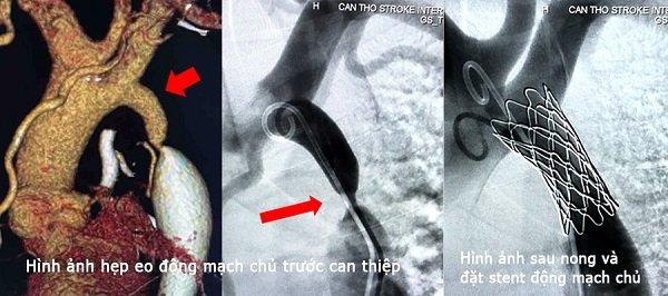
Percutaneous balloon dilation of the aortic coarctation and/or stenting
- Balloon angioplasty is preferred in the cases of re-stenosis and stenosis.
- Aortic stenting in children requires re-intervention to dilate the stent as the child grows. There is now a new generation of stents that can be dilated multiple times as the child grows up.
Complications of angioplasty and stenting through a relatively high waist (15%) include:
- Residual pressure difference (blood pressure between arms and legs remains markedly different)
- Re-stenosis, dissection into aorta
- Aneurysm formation and complications in the femoral artery
8. Monitoring patients with coarctation of the aorta
All patients with coarctation of the aorta who have undergone surgery or dilation should be closely monitored. The aim is to detect and control hypertension and other cardiovascular risk factors. Cardiovascular status should be reassessed annually. Therefore you need:
- Discussion between cardiologists and specialists in adult congenital heart disease. Discussion is done at the first visit to identify risk factors. In particular, anatomical lesions and associated abnormalities will be evaluated more completely.
- Patients following repair of this congenital malformation should be re-evaluated with computed tomography or magnetic resonance aorta in 5 years or less, depending on pre- and post-repair anatomical characteristics.
9. Is coarctation of the aorta dangerous?
Diagnosis of stenosis is not difficult. However, the rate of missed diagnosis is quite high. Therefore, the condition is not treated. The consequences cause many dangerous complications such as:
- Heart failure
- Torn aorta
- Aortic valve stenosis
- Aortic aneurysm
- Infective endocarditis
- Cardiogenic shock, circulatory failure, cardiovascular collapse
- Renal failure due to inadequate perfusion of the kidneys
- Hypertension, especially hypertension in young people
- Coronary artery disease: narrowing of the blood vessels that feed the heart
- Aneurysm in the brain or bleeding in the brain
The best way to prevent coarctation of the aorta is to ensure a healthy pregnancy and not give birth late after the age of 35. As a result, chromosomal abnormalities are minimized.
Although coarctation of the aorta is treatable, careful lifelong monitoring is required as directed by your doctor to detect complications and prevent recurrence. When you have any of the above symptoms in the article, you should go to the doctor immediately for early diagnosis and timely intervention.
Doctor Tran Hoang Nhat Linh
Book an appointment here: h ttps://SignsSymptomsList.vn/