Things to know about periodontal disease in pregnant women

Periodontal disease in pregnant women can cause many complications for both mother and baby. Doctor Truong My Linh will help you learn more about this issue

Many mothers read the ultrasound results of the first prenatal visit and see the word "yolksac". But you don't know what yolksac is? Some moms will question if it's a problem or is the baby developing well? These questions will be answered in the following article.
content
1. What is Yolksac?
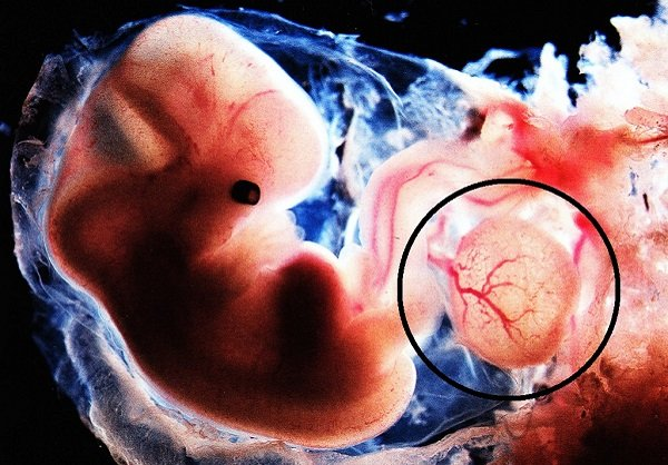
The yolksac, also known as the yolk sac, is a small, membranous structure located outside of the embryo. They have various functions in embryonic development.
The yolk sac is connected to the primitive intestine – the tissue that will later develop into the digestive system – via the yolk sac. Both the yolk sac and the primitive gut are derived from the endoderm.
The yolk sac has many biological functions during early pregnancy, including primitive hematopoiesis and germ cell production.
In terms of pregnancy management, the yolk sac plays an important role in predicting pregnancy outcome through ultrasound imaging.
2. Origin of the yolk sac?
At the end of the second week of pregnancy, after the egg has been released, the sperm has a chance to enter the fallopian tube and combine with the egg to form a zygote.
The zygote continues to divide, moves inside the uterus and develops into a blastocyst. At the 4th week of pregnancy, the blastocyst attaches to the uterine wall and continues to develop.
By the end of the 4th week of pregnancy , the inside of the blastocyst forms 2, and then 3 layers: endoderm, ectoderm, and mesoderm.

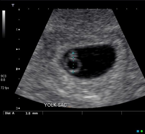
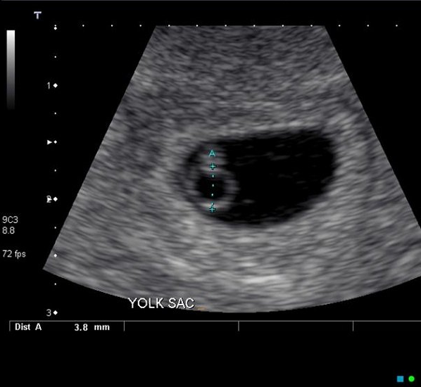
yolk sac image
The yolk sac disappears near the end of the first trimester . Therefore, the yolksac image is not visible on ultrasound from the 14th week of pregnancy onwards.
3. What is the function of the yolk sac?
The yolksac (yolk sac) is responsible for an important part of early pregnancy.
Before the placenta is formed, the yolk sac provides nutrition and metabolism between the mother and the developing embryo.
The yolk sac is also the primary organ of the embryo's blood cell production through blood islets near it. Initial hematopoiesis takes place in the yolk sac. The liver and bone marrow then take over this role, respectively.
Other functions of the yolk sac include:
4. What does the yolksac image on ultrasound mean?
The Yolksac (yolk sac) is significant early in pregnancy because it is the first structure visible on ultrasound.
Doctors can detect yolksac image during transvaginal ultrasound when the fetus is more than 5 weeks old.

Yolksac can be seen on ultrasound when more than 5 weeks pregnant
On transvaginal ultrasound, the gestational sac appears as a circular or oval structure surrounded by a smooth, circular mucosa. In it, the yolksac image appears as a rounder, more translucent structure.
The size of a normal yolksac is from 3 mm to 5 mm, and the normal shape is round. When the yolksac is visible on an ultrasound, it can be confirmed that the fetus is developing inside the uterus.
In addition, images with multiple yolk sacs are also the earliest signs of multiple pregnancy as in the case of twins. The number of yolk sacs matches the number of amniotic sacs if the embryo is alive.
If the yolksac is not visible at 5 weeks, this could be due to: a non-viable miscarriage, or the gestational age may be incorrect. In this case, the doctor will recommend that you repeat the transvaginal ultrasound 1-2 weeks later.
Possible causes of gestational age deviations include:
5. Relationship between abnormal yolksac and miscarriage
Abnormally structured gestational sac and yolk sac are the first signs predicting the possibility of spontaneous abortion. More specifically, an abnormal yolk sac seen on an ultrasound signals a miscarriage that will take place in 7 days, and most often in the first trimester. Therefore, detection of vesicle abnormalities is important in the management of pregnancy and in counseling the mother about possible ongoing miscarriage.
During the first trimester, seeing the yolksac on an ultrasound provides your doctor with important information about your pregnancy outcome. In particular, absence of yolksac or abnormally large or small yolksac is associated with pregnancy loss during the first trimester.
A yolk sac is considered small or large when it is below the fifth percentile or above the 95th percentile corresponding to the expected size. Relatively an abnormal yolk sac is defined as being less than 3 mm in size or larger than 6 mm. Small and large yolk sacs are both correlated with spontaneous abortion. However, this is just a suspicion, not an exact diagnosis.
Other abnormal ultrasound findings associated with miscarriage include an irregularly shaped yolk sac and a calcified yolk sac.
6. Abnormal Yolksac definitely cause miscarriage?
It's important to note that finding an abnormal yolk sac is not always a sign of a miscarriage. There will still be cases where the pregnancy will proceed normally despite the yolk sac being large and irregularly shaped, although uncommon.
After this article, maybe mom also partly understand what yolksac is and the role of yolksac. If the mother is not visible, there is a yolksac at the first prenatal visit. First of all, don't panic and despair. Because the cause may be due to the wrong gestational age calculation. Your doctor will suggest you do a transvaginal ultrasound after 1-2 weeks.
In the case of bad news for the mother, like knowing that the fetus cannot continue to develop. Although it can be disappointing and sad for you, rest assured that early miscarriage is common, often even occurring before a woman even knows she is pregnant. Also, having a miscarriage once doesn't mean you can't get pregnant again.
>> See more: Miscarriage: All the things you need to care about
Periodontal disease in pregnant women can cause many complications for both mother and baby. Doctor Truong My Linh will help you learn more about this issue
The article by Doctor Nguyen Lam Giang about cough during pregnancy is one of the issues that pregnant women feel worried about. Let's find out with SignsSymptomsList.
The yolksac, also known as the yolk sac, is a small, membranous structure located outside of the embryo. There are many different functions in embryonic development.
Doctor Le Ngan Cam Giang's article on whether to drink coconut water during pregnancy is a question many people ask.
Lung maturation injection helps fetal lungs develop more quickly. Learn about the benefits and side effects of this injection with SignsSymptomsList.


