Dyschromia leukodystrophy – what you need to know

Hypochromic leukocytosis is a rare genetic disorder. It is caused by fatty deposits in cells, especially in the brain, spinal cord and peripheral nerves. This buildup is caused by a deficiency in an enzyme that helps break down lipids called sulfates. The brain and nervous system gradually lose function as the myelin sheaths degenerate. Join SignsSymptomsList to learn more about this disease in the following article!
content
- 1. Overview of hypochromic leukocytosis
- 2. How does leukocytosis manifest?
- 3. Causes of hypochromic white matter dystrophy
- 4. Diagnosis of hypochromic leukocytosis
- 5. Treatment methods for hypochromic white matter dystrophy
- 6. Support and care
1. Overview of hypochromic leukocytosis
Hypochromic leukocytosis is divided into 3 types. They are associated with different ages: the late infantile form, the adolescent form and the adult form. Signs and symptoms vary depending on the type. The infantile form is the most common and develops more rapidly than the others.

There is no definitive cure for leukocytosis
There is still no definitive cure for this disease. However, early identification and treatment can help control some of the signs and symptoms and delay the progression of the disease.
2. How does leukocytosis manifest?

Manifestations of hypochromic white matter dystrophy
Deterioration of the myelin that protects nerves leads to a gradual decline in brain and nervous system function, including:
- Loss of sensation, such as touch, pain, heat
- Decreased intelligence, thinking and memory skills
- Difficulty walking, difficulty speaking, difficulty swallowing
- Lose muscle mass
- Diarrhea, urinary incontinence
- Gallbladder problems
- Blindness, hearing loss
- Convulsion
- Emotional and behavioral problems, including emotional instability and substance abuse
Each form of hypochromic leukocytosis occurs at a different age. In addition, they exhibit different initial symptoms and progression rates:
Late infant form
This is the most common form of leukocytosis. The disease occurs in children under 2 years of age. Manifestations: progressive loss of speech and decreased muscle function. Children with this form often have a hard time living past childhood.
Juvenile form
This is the second most common form and begins in children between the ages of 3 and 16. Children often have behavioral and cognitive problems, learning difficulties. Even lost the ability to walk. Although the juvenile form progresses not as rapidly as in the late infant, survival is often less than 20 years after symptoms begin.
Adult form
This form is less common and usually begins after the age of 16. Signs progress slowly. Usually presents with mental problems, drug and alcohol abuse, as well as school and work problems. Psychotic symptoms such as delusions and hallucinations may occur. Adults can survive for several decades after symptoms begin.
3. Causes of hypochromic white matter dystrophy

Hypochromic leukocytosis caused by genetic mutations
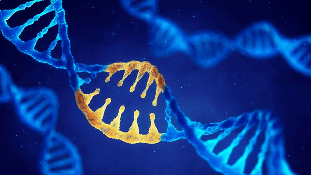
Leukodystrophy is an inherited disorder caused by a genetic mutation. This condition is inherited in an autosomal recessive gene. The abnormal recessive gene is located on one of the undifferentiated chromosomes. To pass it on to their children, both parents must be carriers of the disease, but they often show no signs of the disease. The affected child receives both copies of the abnormal gene – one from the father and one from the mother.
The most common cause of leukocytosis is a mutation in the ARSA gene. This mutation results in a lack of lipid-degrading enzymes called sulfates that accumulate in myelin. More rarely, it is caused by a deficiency in another protein that breaks down sulfates. Found in mutations in the PSAP gene.
The accumulation of sulfates destroys the myelin-producing cells - also called white matter - that protect the nerves. This leads to impaired function of nerve cells in the brain, spinal cord, and peripheral nerves.
4. Diagnosis of hypochromic leukocytosis

Measurement of nerve impulses and muscle and nerve function
Examining and performing some tests to help diagnose as well as assess the extent of the disease.
Blood and urine analysis : Blood tests look for enzyme deficiencies that cause metachromatic leukodystrophy. Urine tests may be done to check for sulfate levels.
Genetic tests : Your doctor may conduct genetic tests for mutations in the genes associated with metachromatic leukodystrophy. They may also recommend testing family members, especially pregnant women (prenatal testing), for mutations in the gene.
Testing nerve signals : Measures nerve impulses and muscle and nerve function by passing a small electrical current through electrodes on the skin. Your doctor may use this test to look for peripheral neuropathy, which is common in people with hypochromic leukocytosis.
Magnetic resonance imaging (MRI) : This method uses strong magnets and radio waves to create detailed images of the brain. They can identify a characteristic tigroid of abnormal white matter in the brain.
Psychological and cognitive tests : Assessment of thinking ability, perception, and behavioral assessment. Mental and behavioral problems may be the first signs in adolescents and adults with leukocytosis.
5. Treatment methods for hypochromic white matter dystrophy
Hypochromic leukocytosis cannot be completely cured. Current treatment is aimed at preventing nerve damage, slowing the progression of the disorder, and preventing complications. Early recognition and intervention can improve the prognosis for some people with this disorder.
Hypochromic leukocytosis can be managed with several treatments:

Medicines can reduce the signs and symptoms of the disease
Medications: Medications can reduce signs and symptoms. Such as behavioral problems, seizures, trouble sleeping, gastrointestinal problems, infections, and pain.
Physical therapy, occupational therapy, and speech therapy: You may have to resort to physical therapy to improve muscle tone, joint flexibility, and maintain as much range of motion as possible. Use a wheelchair, walker, or other assistive device as the illness worsens. Occupational therapy and speech therapy can help improve quality of life.
Nutritional Support : Working with a dietitian can help provide a nutritionally appropriate menu. When dysphagia is severe, the patient may need feeding aids.
6. Support and care

Caring for patients with hypochromic white matter dystrophy
Caring for a child or family member with leukocytosis is truly complex. This requires patience and persistence on the part of both the patient and the family. The level of physical care may increase as the disease progresses. You may not know what to expect, so consider these steps to prepare yourself:
- Find out more information about the disease. Know as much as you can about leukocytosis.
- Genetic counseling if someone in the family has it. This helps you to have better information about these genetic risks to your child.
- Adhere to treatment and schedule follow-up appointments as advised by your doctor.
Above, SignsSymptomsList has provided you with information about hypochromic leukocytosis. The article is for reference only and is not a substitute for medical diagnosis or treatment. If you have any questions, consult your doctor for the best treatment support.
Doctor Nguyen Van Huan