Blood is carried throughout the body through blood vessels. Arteries are blood vessels that carry blood from the heart to other parts of the body. It has a thicker wall, which is stronger and more elastic than a vein. Large arteries branch into small arteries. Finally, the smallest arteries, called arterioles, branch into capillaries. It is where nutrients and wastes are exchanged. These capillaries then combine with other vessels to exit the capillaries to form veins that return blood to the heart.
content
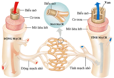
1. Structure and function of the arterial system (arteries)
Structure of arteries
Citadel consists of three layers. In order from outside to inside are: connective tissue layer, smooth muscle layer and endothelial cell layer.
- Connective tissue: Anchor arteries to nearby tissues.
- Endothelial layer: The inside is lined by a smooth tissue called the endothelium.
- Smooth muscle layer: A layer of muscle that allows the arteries to handle the high pressure from the heart.

Structure of blood vessels
The pressure inside the artery is high. Therefore, the artery has a thick vessel wall. Especially, the smooth muscle layer is thick, strong and durable, leading to fast blood flow. Arterioles are the terminal branches of the arterial system. They act as valves that regulate blood flow to the capillaries as needed. This is because the strong muscular layer of the vessel wall can close the vessel lumen, reducing blood flow to the organ or allowing more blood to flow through.
Arterial system
The largest artery is the aorta. This is the main high-pressure conduction vessel that connects to the left ventricle of the heart. The aorta branches into a network of smaller arteries that extend throughout the body. The smaller branches of arteries are called arterioles and capillaries .
Arteries and veins transport blood by two separate vessels: systemic and pulmonary.

Main arteries
Systemic arteries supply oxygen-rich blood to body tissues. Blood returning to the heart through the systemic veins has less oxygen. Since most of the oxygen carried by the arteries has been delivered to the cells.
In contrast, in the pulmonary vessels, the arteries carry oxygen-poor blood to the lungs for gas exchange. The pulmonary veins then carry freshly oxygenated blood from the lungs to the heart to be pumped back into the circulatory system. Although arteries and veins differ in structure and function, they share certain characteristics.
Main function of arteries:
- Helps transport and distribute blood containing nutrients to capillaries throughout the body.
- The arterioles have the function of regulating the distribution of blood into the capillaries.
2. Physiological properties of arteries
2.1. Elasticity
The artery wall is elastic, that is, it is able to expand. When there is an increase in pressure inside the vessel, the vessel will expand and expand according to that pressure. The elasticity of the artery is the physical basis that helps the artery reduce resistance and create continuous blood flow in the artery.
The resistance R of the blood vessel is inversely proportional to the duct radius. Small blood vessels have great resistance, the heart has to work harder to pump blood. In contrast, large blood vessels have small resistance. Blood passes more easily, reducing the work of the heart to pump blood. When a vascular segment has changes in pressure, thanks to the elasticity of the blood vessels will be dilated and increased in radius. This reduces the resistance and helps the heart to pump blood.

Arterial elasticity helps blood circulation
In the aorta, the heart contracts to produce intermittent blood flow with each systole. The pressure when the heart contracts, partly pushes blood to move in the aorta, partly dilates the artery wall. During diastole, the heart does not push blood, but the elastic artery wall contracts again, forcing blood to continue to circulate to the periphery.
Therefore, the elasticity of the artery converts the intermittent intermittent blood flow at the origin of the aorta into continuous blood flow, which is smoother and more regular to the periphery. The smooth blood flow regime at the end of the arterioles is consistent with the peripheral tissue nourishing blood supply.
The elasticity of the vessel walls decreases with age. The elastic properties of the aortic wall can be clearly determined when examining the relationship between pressure and volume in the aorta.
2.2. Contractility
The arterial wall contains smooth muscle. Therefore, it can actively change diameter, especially in arterioles. When the smooth muscle fibers of the vessel wall contract, the vessel volume decreases and the vessel pressure increases. When vascular smooth muscle fibers relax, vessel volume increases and pulse pressure decreases.
3. Pulse phenomenon
Pulse palpation is the feeling of bouncing pulse at the tip of the finger when placing the hand lightly on the artery. During systole, pressure from the heart not only pushes blood forward, but also causes pressure waves to propagate along the artery. The pressure wave stretches the artery wall as it passes. We can feel it and call it a pulse.
The propagation rhythm of the pressure wave is independent and higher than the velocity of the blood flow. Approximately 4 m/s in the aorta; 8 m/s in the great artery and 16 m/s in the small artery in young adults. Thus, a palpable wrist pulse occurs 0.1 s after the peak of the pumping phase at ventricular contraction.
As you age, the walls of your blood vessels become stiffer, so pulse waves move faster.
4. Arterial blood pressure
4.1. Define
Arterial blood pressure is the force exerted by the blood on a unit area of the artery wall. Blood flow in an artery is the result of two opposing forces: the force of the heart pushing blood and the resistance of the artery wall. In it, the blood pushing force of the heart is stronger. Therefore, blood flows through the arteries at a certain speed and pressure.
The unit of blood pressure is: mmHg or Kpa (KiloPascal). 1 KPa = 7.5 mmHg.

Blood pressure
4.2. Parameters of blood pressure
Systolic blood pressure, also known as maximum blood pressure. It is the upper limit of periodic fluctuations in blood pressure in the pulse. It represents the pumping power of the heart.
>> Learn more related information: Is isolated systolic hypertension dangerous?
Diastolic blood pressure, also known as diastolic blood pressure. It is the lowest limit of cyclic fluctuations of blood pressure in the pulse. It represents the resistance of the vessel wall.
It is the difference between the maximum and minimum blood pressure. The pressure depends on: the force of contraction of the heart and the resistance of the blood vessels from the heart to the capillaries.
Is the average of all blood pressures measured over a period of time. Average blood pressure represents the actual work of the heart. The pressure that produces a continuous flow of blood with a flow equal to cardiac output. Mean blood pressure is closer to diastolic pressure than systolic pressure during the cardiac cycle.
Mean blood pressure = diastolic pressure + 1/3 of blood pressure.
4.3. Physiological changes in blood pressure
As you age, blood pressure increases. The degree of hypertension parallels the degree of arterial stiffness. That is, increased diastolic blood pressure. Then there is an increase in systolic blood pressure.
With normal blood density, in the upright position, the mean blood pressure in the transcardiac artery is 100 mmHg. Due to the influence of gravity, if the artery is 1 cm higher than the heart, the blood pressure will decrease by 0.77 mmHg. When the artery is 1 cm lower than the heart, the blood pressure increases by 0.77 mmHg.
Eg:
The mean blood pressure of the great artery at the head, 50 cm from the heart is: 100 - (0.77 x 50) = 62 mmHg.
The average blood pressure of the great artery in the leg, 105 cm from the heart is: 100 + (0.77 x 10⁵) = 180mmHg.
Eating too much salt causes blood pressure to rise. Eating a lot of meat, blood pressure increases because more protein in the blood increases viscosity.
>> You need to pay special attention to your diet to maintain a stable blood pressure level. People with high blood pressure need a separate diet. Read more articles Diet for people with high blood pressure: How reasonable?
When exercising, in the early stages, blood pressure increases due to many emotional reflexes before exercise. Then the blood pressure gradually decreases, but remains higher than normal. Hard work, low blood pressure is a sign that the heart cannot meet the needs and is not effective enough to complete the function of pumping blood.

Blood pressure will be affected when you exercise
4.4. Method of measuring blood pressure
4.4.1. Direct measurement
Insert the catheter into the artery, measure blood pressure with a mercury sphygmomanometer (Ludwig).
4.4.2. Indirect measurement
Measure arterial blood pressure by indirect method: Compress a relatively large diameter artery (usually brachial artery) with an air bag. Inflate the air bag to create air pressure, from which to measure the air pressure in the air bag and deduce the arterial blood pressure with a sphygmomanometer. There are two methods: pulse and listen.
When the air bag is not inflated, it is normal to feel the pulse when touching. Inflate the air bag until the pressure in the cuff is greater than the systolic blood pressure. The artery is completely compressed, blood cannot flow through, so it can no longer catch the pulse.
Continue to inflate by 30 mmHg and then begin to deflate the airbag until the airbag pressure is equal to and below the systolic blood pressure. The artery is no longer compressed, blood can flow through the compressed area, so the pulse is felt again, corresponding to the systolic blood pressure. The pulse is then still felt as the pressure in the airbag continues to decrease until 0 mmHg. Therefore, pulse measurement only indicates systolic blood pressure, not diastolic blood pressure.
Blood pressure is usually measured by auscultation. Use the flat side of the stethoscope to rest on the brachial artery 2 cm above the elbow crease.
When the air bag is not inflated, no sound is heard when the stethoscope is placed over the brachial artery. When the air bag is inflated, the narrowing blood vessels will make a noise. Until the pressure in the air sacs is greater than the systolic blood pressure, the artery is completely compressed and no sound is heard.

Measuring blood pressure by listening method
Continue to inflate by 30 mmHg and then begin to deflate the airbag until the pressure in the airbag equals the systolic pressure in the artery. Blood breaks through the blockage during systole, bouncing back into the quiet column of blood below, causing the first sound, which is the systolic pressure. As the pressure in the airbag continues to decrease, a noise is heard each systole, which gets louder, then decreases, and then disappears altogether. It was the Korotkoff noise.
The pressure reading at the time of sound loss is the diastolic blood pressure. Korotkoff sounds are caused by blood vortexing in the brachial artery.
5. Possible problems in the arteries
The accumulation of cholesterol (a waxy substance) in plaques called plaques in the artery walls. These plaques can become fragile, leading to heart-related complications. Atherosclerosis in the arteries of the heart, brain, or neck can lead to heart attacks and strokes.

This is a dangerous situation
Arteritis, which may involve one or more arteries at once. Most vasculitis is caused by an overactive immune system.
Loss of vision in one eye due to temporary loss of blood flow to the retina (light-sensitive tissue located at the back of the eye). It usually occurs when part of the cholesterol plaque in one of the carotid arteries (the arteries on the sides of the neck that supply blood to the brain) ruptures and travels to the retinal artery (the artery that supplies blood and nutrients to the retina). ).
Narrowing of the arteries, often due to atherosclerosis. When narrowing occurs in the arteries of the heart, neck, or legs, the limitations in blood flow can cause serious health problems.

Blocked arteries in the legs
Atherosclerosis causes narrowing of the arteries in the legs or groin. Restricted blood flow to the leg can cause pain or poor wound healing.
It occurs when a blood clot suddenly forms in one of the arteries, blocking blood flow. Immediate treatment is needed to restore blood flow in the artery.
The condition occurs when there is a sudden blood clot in one of the arteries that supply blood to the heart.
The condition occurs when there is a sudden blood clot in one of the arteries that supply blood to the brain. A stroke can also happen when one of the arteries in the brain bursts, causing bleeding.
Inflammation of the temporal artery in the scalp. Jaw pain when chewing and pain on the scalp are common symptoms.
Atherosclerosis is associated with narrowing of the arteries that supply blood to the heart muscle. Coronary artery disease makes a heart attack more likely.
Atherosclerosis with stenosis of one or both carotid arteries in the neck. Carotid artery disease also makes stroke more likely.
6. Methods for examining arteries
A thin, flexible tube is inserted into the artery. Then a special dye is injected and an X-ray shows blood flow through the artery. Areas of narrowing or bleeding within an artery can often be identified through angiography.
Use a CT scanner to take multiple tomography sheets and use a computer to create detailed images of the arteries. A CT-A scan can often show narrowing or other problems in an artery with less risk than a conventional angiogram.

Tomography machine
Whether it's exercise or medication, the heart will be stimulated to beat faster. As this stress increases blood flow through the heart, narrow spots in the coronary arteries can be identified through various testing techniques.
An MRI scanner uses high-powered magnets and a computer to create highly detailed images of structures inside the body. The MRA is a setting that allows the MRI scanner to best display the images of the arteries.
A catheter (thin, flexible) is inserted into one of the arteries in the groin, neck, or arm and inserted into the heart. A contrast-enhancing dye is injected through the catheter so that blood flow through the coronary artery can be seen on the X-ray screen. The blockage in the artery can then be found and treated.
A small piece of the artery is removed and examined under a microscope. Usually to diagnose vasculitis. The temporal artery of the scalp is most commonly biopsied.
Arteries are an important structure of the vascular system as well as of our body. It plays a role in distributing blood from the heart to other organs to nourish the body. However, they are also prone to some problems such as atherosclerosis, blockage, inflammation ... affecting health, even life. Therefore, we must regularly check our health and maintain a healthy lifestyle, to avoid problems related to the arteries.
Doctor Truong My Linh