The structure and function of the eye is quite complex. The main function of the eye is to regulate the amount of light entering the eye and help us see objects clearly, whether near or far. Humans can see because the eyes are constantly creating images and rapidly transmitting signals to the brain.
The eye socket is the bony cavity that contains the eyeball, muscles, nerves, blood vessels, and other structures that help produce and drain tears. The eye sockets are usually pear-shaped.
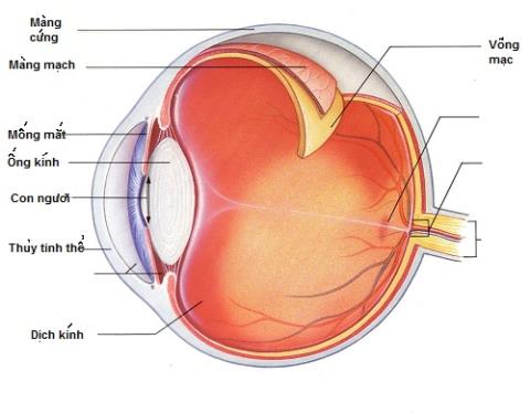
Outside the eyeball is a relatively hard, white layer called the sclera (white).
Near the front of the eye, the sclera is covered by a thin, transparent membrane called the conjunctiva, which runs to the edge of the cornea. The conjunctiva also covers the moist surface of the eyelid and eyeball.
Light enters the eye through the cornea, the membrane in front of the iris, and the pupil. The cornea protects the eye and acts as a lens to help focus images onto the retina. Thanks to that, we can see the image.
After passing through the cornea, light will pass through the pupil (the black round hole in the center of the eye).
The iris - the pigmented membrane that surrounds the pupil - helps balance the amount of light entering the eye. The iris allows more light to enter the eye (by dilating the pupil) when the environment is dark and allows less light to enter the eye (by constricting the pupil) when the environment is bright. The mechanism of the pupil dilation resembles the aperture of a camera lens when the light of the environment changes. The size of the pupil is controlled by the sphincter and dilator muscles of the pupil.
Behind the iris is the lens. By changing shape, the lens helps focus light onto the retina. Through the action of small muscles (the ciliary body), the lens becomes thicker to see near objects clearly and thinner to see distant objects clearly.
The retina contains light-sensitive cells (photoreceptors) and blood vessels to nourish them. The most sensitive part of the retina is the macula, where there are millions of layers of photoreceptors (especially cones). Because the density of these cells present in the macula is very high, the images we see also become more detailed. People often compare cone density to megapixels in digital cameras, the more cells, the higher the resolution.
Each photoreceptor is associated with a nerve fiber. Nerves originate from photoreceptors that are bundled together to form the optic nerve. The optic disc, the beginning of the nerve of the same name, is located behind the eye.
Photoreceptors convert images into electronic signals that are sent to the brain by the optic nerve. There are 2 types of photoreceptors: rods and cones.
Cones are responsible for the detail, sharpness and color of the image. These cells are concentrated mainly in the macula.
Rod cells are responsible for peripheral vision and in low light conditions. Compared to cones, these cells are more numerous in number and also more sensitive to light. However, they are not color-specific and support image detail like cones. Rod cells are concentrated mainly in the periphery of the cornea.
The eyeball is also divided into two parts, each of which is filled with vitreous fluid. The pressure created by the fluid helps the eyeball to maintain its original shape.
The anterior part (anterior chamber of the eye) extends from the inside of the cornea to the anterior surface of the lens and is filled with aqueous humor, which nourishes the structures inside the eye. The front chamber is divided into 2 compartments. The anterior chamber extends from the cornea to the iris. The posterior chamber extends from the iris to the lens. Normally, aqueous humor is produced in the posterior chamber, moving slowly from the pupil to the anterior chamber. The fluid then exits the eyeball through channels located where the iris meets the cornea.
The posterior chamber of the eye extends from the back of the lens to the retina. It contains a jelly-like liquid called the vitreous.

Image of the structure of the eye
Trace the visual conduction pathway
Nerve signals travel from the eye, along the corresponding optic nerve and other nerves (called the optic nerve pathway) to the back of the brain, where images are interpreted. The two optic nerves meet at the optic nerve, which is the area behind the eye, just in front of the pituitary gland, and just below the front part of the brain (the cerebrum). There, the optic nerve in each eye is divided, and half of the nerve fibers from each side cross to the other side and continue on to the back of the brain. Therefore, the right brain receives information from both optic nerves for right vision and the left brain for left vision. The central part of the vision that is often overlapped is seen by both eyes (called binocular vision).
Each eye will observe the object from many different angles. Therefore, the information the brain receives from the eyes also differs, although the information often overlaps. The brain interprets the information to create a complete image.