Human seminal vesicles: structure and function

The seminal vesicles in humans are part of the male reproductive system. This unit has a distinct structure and function. It plays a major role in semen production. So what is the structure of the seminal vesicles? How is the function? What are the common diseases? The following article will help readers find answers to these questions.

content

1. What is the human seminal vesicle?

The seminal vesicles are also known as the seminal vesicles. This is a pair of glands located in the male pelvis. They function to produce many of the components that make up semen. After all, they provide about 70% of total semen.

2. Characteristics of human seminal vesicles

Each seminal vesicle is pyramidal in shape, with a length of about 5 cm. The diameter of each seminal vesicle is 3-4cm and they are 10cm long when unrolled. The upper sides of the seminal vesicles are covered by the peritoneum. The base of each bag points upwards as well as towards the back.

See more articles on the same topic:  What to do when itchiness of the tip of the penis?

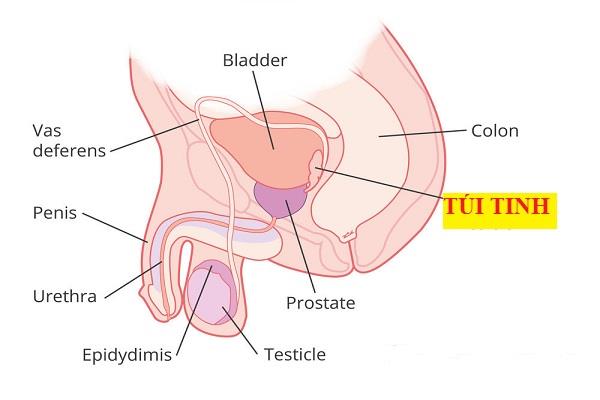
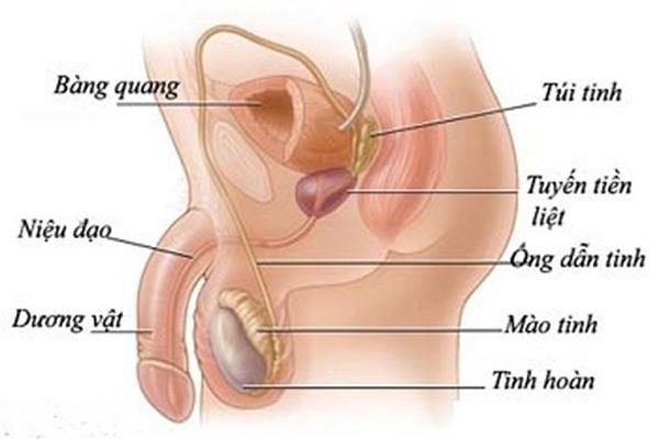
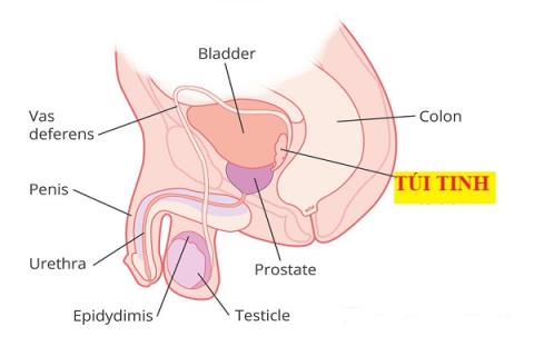
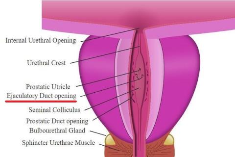
At the lower end, each sac narrows to form a straight duct that communicates with the vas deferens. The vas deferens carry sperm. When the seminal vesicles combine with the vas deferens, the ejaculatory duct is formed. This duct then opens into the urethra of the prostate at an opening called the mound.

Human seminal vesicles: structure and function

The seminal vesicles in men.

3. Location of seminal vesicles

The anterior surface of each seminal vesicle is in contact with the posterior wall of the bladder. However, the back of each pouch is separated from the rectum by the rectal wall or Denonvillier fascia. Located below the seminal vesicles is the prostate gland. The ureter is located in front of the seminal vesicles. In the center of the seminal vesicles are the ducts of the vas deferens, while the veins of the prostatic plexus are adjacent to them.

4. Structure of seminal vesicles

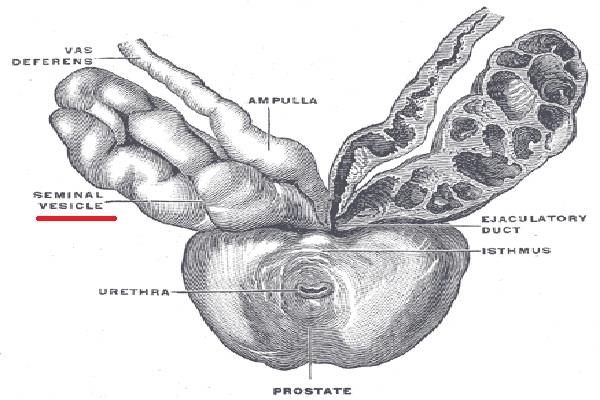
An individual seminal vesicle consists of a single coiled tubule in which there are several branching vesicles. The tubules of the seminal vesicles are made up of three different layers:

  • Specialized cells responsible for the production of seminal vesicles are located in the inner layer.
  • Smooth muscle tissue is located in the middle layer.
  • The outer layer is the connective tissue layer.

Part of the seminal vesicles and vas deferens join together to form the ejaculatory duct, which eventually drains into the prostatic portion of the urethra. During ejaculation, the smooth muscle layer of the seminal vesicle contracts, releasing seminal fluid into the ejaculatory duct.
Human seminal vesicles: structure and function

Anatomical structure of seminal vesicles.

5. Blood vessels and nerves

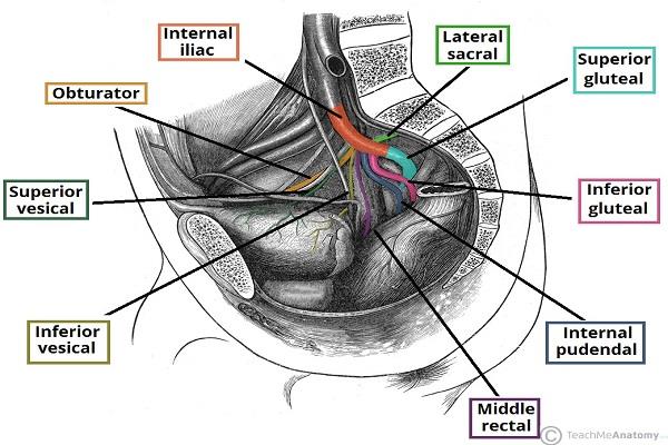
The arteries supplying the seminal vesicles are branches of the inferior cystic artery and the middle rectum. They all arise from the internal iliac artery. The parasympathetic nervous system of the seminal vesicles originates from the hypogastric plexus. Sympathetic supply is from the superior lumbar and hypogastric nerves.

Human seminal vesicles: structure and function

Blood vessels in the seminal vesicles.

6. Histology

Under low magnification, the lumen appears honeycomb due to the irregular lumen and presence of diverticula. The wall of the seminal vesicles consists of three main layers. The outer layer consists of connective tissue, while the middle layer consists of smooth muscle. Inside there is also the appearance of a mucosal layer.

See also:  What are the functions of the scrotum?

The middle layer of smooth muscle is arranged in two layers: an outer longitudinal layer and an inner circular layer. Both of these layers are supplied by the sympathetic nervous system. The inner layer is composed of pseudostratified columnar epithelium and also contains secretory cells. These secretory cells have lipid droplets in their cytoplasm that give a foamy appearance.

Epithelial cells have some variation in size and shape and may often contain brown lipofuscin granules. Although the seminal vesicles do not store sperm, sperm are usually seen under a microscope. It is also thought to enter the seminal vesicles by regurgitation through the vas deferens.

7. Embryology

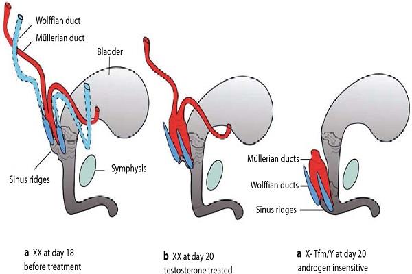
The seminal vesicles develop from the seminiferous or Wolffian ducts. These ducts originate in the mesoderm, the middle layer of one of the three primary germ layers in the embryo. The other two layers are ectoderm and endoderm. The seminal vesicles are formed from the budding of the distal fascia at around 10-12 weeks' gestation.
Human seminal vesicles: structure and function

The vas deferens.

8. Function of the seminal vesicles in humans

The function of the seminal vesicles is to both produce and store fluid. That fluid eventually becomes semen. This fluid covers about 70% of semen. Reliable source of fluid secreted during ejaculation.

The fluid produced in the seminal vesicles provides an environment that is essential for the normal functioning and survival of sperm. The main components of this fluid are:

  • Fructose: A type of sugar that provides energy for sperm
  • Alkaline fluid, which helps to neutralize the acidity of the male urethra and female vagina.
  • Proteins, such as semenogelin, form a protective gel-like layer around the sperm.
  • Phosphorus and potassium, which help sperm move
  • Prostaglandin, a hormone that plays a role in reducing a woman's immune response to semen.

Human seminal vesicles: structure and function

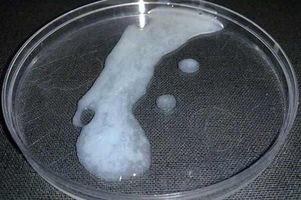
The seminal vesicles play a major role in the production of semen.

9. Possible diseases in the seminal vesicles

Human seminal vesicles can be affected by the following diseases:

9.1. Infections and abscesses of the seminal vesicles

A seminal vesicle abscess occurs when bacteria enter the seminal vesicles. This can happen after an infection of the urethra or prostate gland. Treatment for a seminal vesicle infection is with antibiotics.

Human seminal vesicles: structure and function

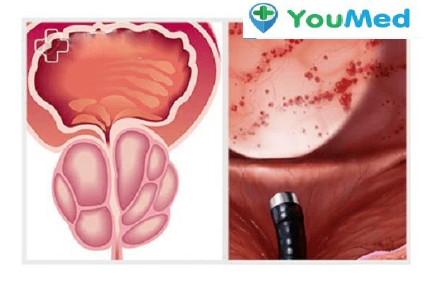
Inflammation of the seminal vesicles.

In some cases, a pocket of pus, called an abscess, can result from an infection. In this case, the most practical treatment is antibiotics combined with drainage of the abscess.

9.2. seminal vesicles

Seminal vesicles are usually asymptomatic, which means they are not apparent. They can be congenital or acquired. Acquired seminal vesicle cysts can arise from conditions such as scarring from an infection or previous prostate surgery. Depending on the size of the seminal cyst, laparoscopic surgery may be performed to remove it.

Human seminal vesicles: structure and function

Seminal vesicle cyst.

9.3. Seminal vesicles

The presence of stones in the seminal vesicles is very rare. They are thought to form due to inflammation or structural abnormalities in the seminal vesicles. Reflux of urine back into the ejaculatory duct can also lead to stone formation. The seminal vesicles need to be removed. Especially when there are many stones or large stones. This can be done through a laparoscopy or laparoscopy.

Human seminal vesicles: structure and function

Seminal vesicle stones cause lower abdominal pain.

9.4. Testicular cancer

Cancer occurring in the seminal vesicles is very rare. As of 2000, there were only 48 confirmed cases. The cause of cancer in the seminal vesicles is still unknown.

Human seminal vesicles: structure and function

Testicular cancer.

Many cancers involving the seminal vesicles occur as a result of invasion of the seminal vesicles by another malignancy. An example is prostate cancer. The proximity of the seminal vesicles to the prostate makes this invasion more likely.

9.5. When to see a doctor?

When the following symptoms appear, we should see a doctor immediately:

  • Pelvic pain or pain in the penis
  • There is pain or discomfort during ejaculation.
  • Blood in the ejaculate.
  • The amount of semen when ejaculating is less than normal.
  • Urinary disorders such as: frequent urination, painful urination, hematuria, pyuria, etc.

Human seminal vesicles: structure and function

Doctor specializing in Orthopedics.

9.6. Tests that support the diagnosis of pathology in the seminal vesicles

In addition to a physical exam, specialists may order a number of tests to aid in the diagnosis. The most typical tests include:

  • Fresh semen analysis to detect white blood cells.
  • Semen culture can detect bacteria that cause seminal vesicle infections.
  • Ultrasound of the seminal vesicles through a rectal probe. Or an abdominal ultrasound with a full bladder. Both of these methods make it easy to visualize the seminal vesicles.
  • A semen analysis can be done with a semen sample. The level of fructose in semen is a measure of the function of the seminal vesicles. Accordingly, low or no fructose levels may indicate a blockage inside the seminal vesicles.

Human seminal vesicles: structure and function

Translate map.

10. Measures to have a healthy seminal vesicles

Since the seminal vesicles play a major role in the fluid present in semen, it is important to keep the seminal vesicles healthy. Follow the tips below to have good sperm and maintain reproductive health.

10.1. Safe sex activities

Make sure to practice safe sex with any new sexual partners. Condoms not only prevent unwanted pregnancy, but they can also prevent sexually transmitted infections. Infection can lead to inflammation and scarring of the urogenital tract, including the seminal vesicles.

Human seminal vesicles: structure and function

Safe sex activities.

10.2. Try to maintain a reasonable weight

Try to maintain a healthy weight so that your BMI is between 18.5 and 23. A BMI of more than 23 has been associated with delayed sperm motility as well as lower sperm count. .

Human seminal vesicles: structure and function

Maintain a healthy weight.

10.3. Have a scientific and healthy diet

You should maintain a diet rich in vegetables and fruits. Because these foods contain antioxidants, it is very beneficial for the health of the seminal vesicles and sperm. We should prioritize whole grains and lean meats. Avoid processed foods. Limit your consumption of foods high in saturated fat and sugar.

Human seminal vesicles: structure and function

Whole grains.

10.4. Quit smoking habit

Smoking can make your sperm less mobile. It also reduces your sperm count. Quitting the habit of smoking can be difficult, but it is possible. Your doctor can help you create a quit plan that's right for your particular situation.

See also:  How to detox from masturbation that everyone should know .

10.5. Don't ignore worrisome symptoms

If you experience any symptoms that are different than usual, you should see your doctor right away. It is best to choose medical facilities that specialize in andrology. Here, doctors will examine and order tests. Finally, make a definite diagnosis and have a specific treatment direction.

See more:  Pimples on the testicles are a sign of what disease?

In short, the human seminal vesicles play a very important role in the formation of semen. It also plays an important role in male fertility. Therefore, to protect the reproductive function, men should protect the health of the seminal vesicles. As well as go to the doctor immediately when there are abnormal symptoms in the locations related to the seminal vesicles.

Dr. Nguyen Lam Giang

Vaginal pH and what you need to know

Vaginal pH and what you need to know

Article by Doctor Nguyen Thanh Xuan about Vaginal pH - the vagina has a very rich resident microflora including beneficial and harmful microorganisms

The kneecap: Structure and function

The kneecap: Structure and function

On the human body, there are about 49 sesame bones of different sizes. Sesamoid bones are small bones located below tendons, commonly found in the joints of the hands and feet.

Human seminal vesicles: structure and function

Human seminal vesicles: structure and function

The seminal vesicles in humans are part of the male reproductive system. This unit has a distinct structure and function. It plays a role in semen production

What is the function of the ureters in your body?

What is the function of the ureters in your body?

The article was consulted by doctor Nguyen Huynh Uyen Tam about ureters, ureteral function and some related diseases.

Testicles: Basic Knowledge

Testicles: Basic Knowledge

Article by Doctor Nguyen Doan Trong Nhan about testicles. There are many conditions that affect the testicles and can sometimes lead to infertility.

Enamel: The strongest structure in the body!

Enamel: The strongest structure in the body!

Enamel is the outermost layer, plays a protective role and is also the strongest component. Although hard, enamel can still crack, chip and be dissolved by acids

Gums: Important soft tissue that surrounds teeth

Gums: Important soft tissue that surrounds teeth

Gums play an important role in the oral system. It helps to cover, create aesthetics for the mouth area, protect and prevent the penetration of bacteria

The temporal lobe: Anatomy and function

The temporal lobe: Anatomy and function

The temporal lobe is one of the main lobes that make up the human brain. This lobe of the brain has distinct functions.

The lacrimal gland: Anatomical and functional features

The lacrimal gland: Anatomical and functional features

Doctor Nguyen Lam Giang's article about the lacrimal gland, a very specific exocrine gland in humans, thanks to the lacrimal gland we can express our emotions

The tibia: location, structure and function

The tibia: location, structure and function

Article by Doctor Luong Sy Bac about tibia. The tibia is the large bone in the lower leg that bears the main force of the body and is often injured.