The occipital lobe in the human brain is one of the components that make up the brain. It has distinct anatomical and functional features. Damage to this lobe will significantly affect the functions of the body. So what is the anatomical structure and function of this lobe? What diseases are associated with this lobe? Let's go with SignsSymptomsList to find the answer through the following article.
content
1. What is the occipital lobe?
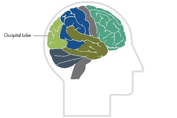
The occipital lobe or occipital lobe is one of the four main lobes of the cerebral cortex in the mammalian brain. The occipital lobe is the visual processing center of the mammalian brain, containing most of the anatomical regions of the visual cortex. The primary visual cortex is Brodmann's area 17, commonly referred to as V1 (vision one).
Occipital lobe
The human V1 region is located medial to the occipital lobe in the fissure. The full extent of V1 usually continues into the occipital pole. V1 is often referred to as the striated cortex because it can be identified by a large band of myelin. The areas of visual control outside of V1 are called the external cortex.
See also: What do you know about temporal lobe epilepsy?
There are many peripheral regions and these are specialized for different visual tasks. Such as visual spatial processing, color discrimination and motion sensing. The name derives from the upper occipital bone, which is named after the Latin. Bilateral damage to the occipital lobes can lead to cortical blindness.
2. Anatomical structure of the occipital lobe
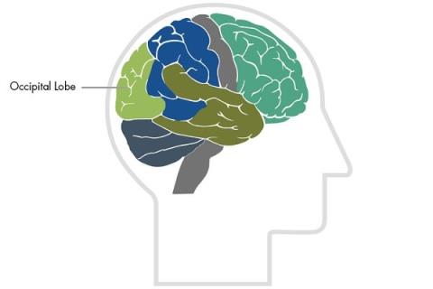
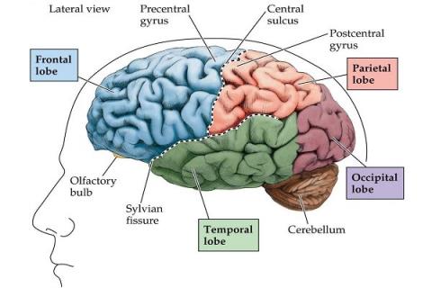
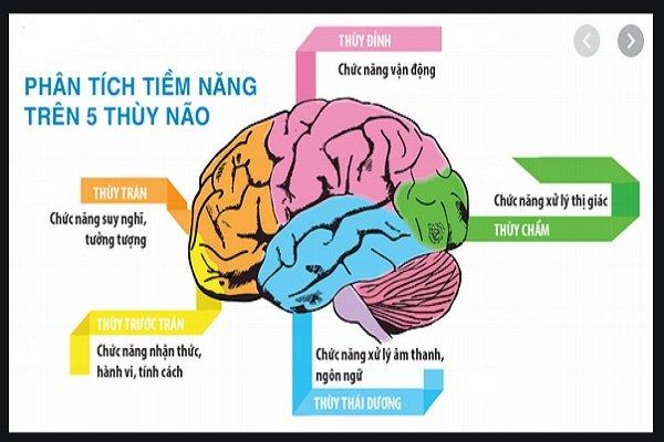
The two occipital lobes are the smallest of the four paired lobes in the human brain. Located at the back of the skull, the occipital lobe is part of the hindbrain. The lobes of the brain get their name from the bones above and from the occipital bone that overrides the lobes.
The lobes are located above the cerebellar tentacle, a structure of the dura mater that separates the cerebrum from the cerebellum. They are structurally isolated in their respective cerebral hemispheres by cleft palate detachment. At the anterior edge of the occipital lobe are several occipital gyrus, which are separated by the lateral occipital fissure.
The lobes of the brain
The occipital lobes are located in the posterior part of the upper brain. They are located behind the temporal and parietal lobes and above the cerebellum. Separated from the cerebellum by a membrane called the tentacle.
The surface of the occipital lobe is a series of folds, including ridges known as the gyrus. And the depressions are called grooves. Because there is no ordered structure to the occipital lobe, the scientists used grooves and gyrus to define the region of the lobe.
The occipital lobe itself contains different sections or regions, and each of these regions has a different set of functions. Consists of:
- Boundary lateral organs.
- Language.
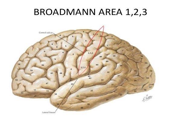
- The primary visual cortex, known as Brodmann's area 17 or V1.
- The secondary cortex, known as Brodmann's area 18 and 19 or V2, V3, V4, V5, surrounds the primary visual cortex.
- Back thread.
Broddmann's brain region
3. Function of the occipital lobe
Researchers once thought that the occipital lobe controlled only visual functions. But in recent years, they have discovered that certain parts of this lobe receive input from other brain regions.
Specifically, an area of the brain called the dorsal region receives input from brain regions involved in vision. Accompanied by regions unrelated to visual processing. This suggests that the occipital lobe may perform additional functions. Or researchers haven't identified all the regions of the brain involved in visual processing.
Although we know that the occipital lobe is dedicated to vision, the process is complex and involves several distinct functions.
These include:
- Mapping the visual world, which helps with both spatial reasoning and visual memory. Most vision involves some kind of memory, since scanning the visual field requires you to recall something you saw just a second ago.
- Specifies the color property of the items in the visual field.
- Assess distance, size and depth.
- Recognizes visual stimuli, particularly familiar faces and objects.
- Transmits visual information to other brain regions so that those lobes can encode memories. Simultaneously assign meaning, generate appropriate linguistic and motor responses.
- Constantly responding to information from the world around.
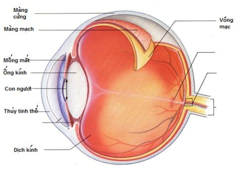
- Receive raw image data from perceptual sensors in the retina of the eye.
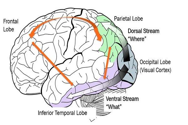
Visual function
4. Primary visual brain training
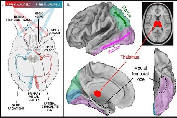
The primary visual cortex, known as Brodmann area 17 or V1, receives information from the retina. It then interprets and transmits information regarding the space, position, motion, and color of objects in the visual field. It does this through two different pathways known as the ventral stream and the dorsal stream.
See also: Generalized epilepsy and what you should know
5. Secondary visual brain
Secondary visual cortex – known as Brodmann area 18 and 19 or V2, V3, V4, V5. It receives information from the primary visual cortex. The secondary cortex processes many of the same types of visual information.
6. Abdomen
The ventral flow is a pathway that the primary visual cortex uses to send information. It delivers information to the temporal lobe, which interprets information and helps the brain provide meaning to objects in sight. This helps with object recognition and gives a conscious perception of what a person is seeing.
See more: Brain tumor: Is it really an incurable disease?
7. Backflow
The dorsal stream is another pathway that the primary visual cortex uses to send information. It shares information about an object's position and carries it to the parietal lobe. Acquire other spatial and geometrical information about objects in the field of view.
8. Other Contributions
Modern science has revealed much about the way in which the occipital lobe reveals the visual world. However, researchers are still learning new information about the occipital lobe and how exactly it works.
No part of the brain is truly independent, and this includes the occipital lobe. For example, the occipital lobe takes information from the retina in the eye and transfers it to the visual world. As such, it depends a lot on the eyes themselves.

Eyes play an important role in visual function
The eyes themselves have muscles to control. The motor cortex in the brain is responsible for these movements, so also plays a role in vision.
The temporal and occipital lobes also have important interactions. The temporal lobe gives meaning to visual information interpreted from the occipital lobe. It also stores information, to a degree, in the form of memories. In some cases, other parts of the brain can also compensate for any damage affecting this lobe.
9. How does the occipital lobe interact with other areas of the body?
No part of the brain is an independent organ that can function without information from other parts of the body. The occipital lobe is no exception. Although its main role is to control vision, damage to other brain regions and body parts can inhibit vision.
See also: What to know about Peripheral nerve damage
Furthermore, some evidence suggests that, when the occipital lobe is damaged, neighboring brain regions can compensate for some of its function. The occipital lobe is highly dependent on:
- The eyes, especially the retina, receive and process visual information, which is then further processed by the occipital lobe .
- The frontal lobe, which contains the motor cortex of the brain. Without motor skills, the eyes cannot move or receive information from the surrounding areas.
- The temporal lobe, which helps assign meaning to visual information, in addition to encoding it into memory.
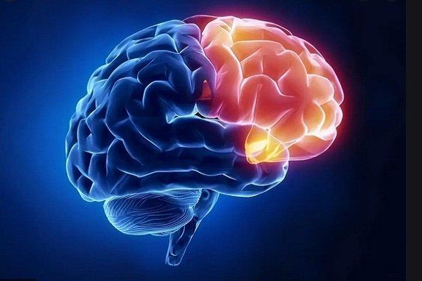
The frontal lobe also affects the occipital lobe
10. Medical conditions associated with the occipital lobe
Dysfunction in the occipital lobe can lead to one or more dysfunctions in the brain, vision, or daily function. It may cause or contribute to any of the following conditions.
10.1. Blind
Because the occipital lobe is involved in vision, one possible outcome of damage to this area is total or partial blindness. However, vision loss is not always straightforward. And instead, the person may lose one or more specific functions of vision.

Blind
10.2. Anton's syndrome
Anton syndrome is a rare form of blindness that occurs without the patient even knowing it. They may deny their vision loss of vision. Even when a healthcare professional presents evidence that they have lost their vision.
Anton's syndrome
10.3. Riddoch's syndrome
Riddoch syndrome is a rare condition in which a person can only see moving objects. Static objects do not appear in their field of vision. The person is also unable to perceive shapes or colors.
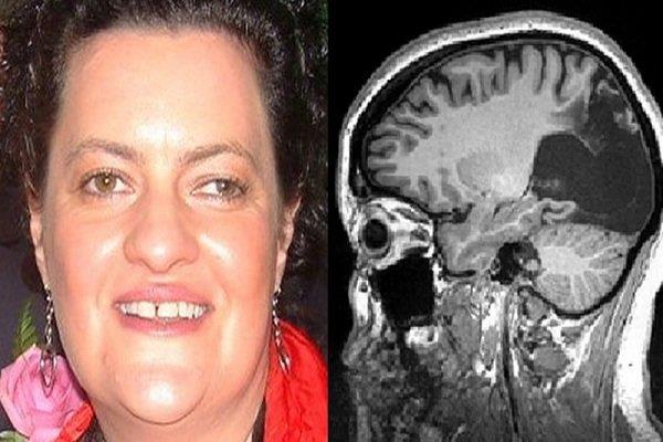
Riddoch's syndrome
10.4. Epileptic
In some cases, epilepsy has been associated with the occipital lobe. A person is prone to having occipital seizures or seizures due to sensitivity to light. When this happens, flashing lights or colorful images can cause these seizures.
10.5. Other types of dysfunction
The type of dysfunction that affects the body can vary depending on where the dysfunction or damage occurs in the occipital lobe. Some examples may include:
- Difficulty recognizing everyday objects
- Understanding basic colors, shapes, or sizes becomes difficult.
- The recognition of familiar faces is difficult.
- Difficulty balancing, moving or standing.
- Visual hallucinations, such as flashes of light.
- Change in depth perception.
- Difficulty detecting moving objects.
- Difficulty reading or writing due to difficulty in word recognition.

Difficulty maintaining body balance
11. Conclusion
The occipital lobe is one of the four major lobes in the mammalian brain. This is the lobe primarily responsible for interpreting the visual world around the body. Such as the shape, color and position of an object. It then relays this information to other parts of the brain. The places give this visual information its meaning.
Dysfunction in the occipital lobe can cause a number of bodily dysfunctions. For example, uneven vision, difficulty standing, and blindness. Some conditions, such as epilepsy, may also be associated with dysfunction in the occipital lobe.
Dr. Nguyen Lam Giang
