The umbilical cord is like a long tube containing blood vessels connecting the mother and the baby through the placenta. The main function of the cord is to transport oxygen and nutrients to the baby. In this article, we will discuss the function and structure of the umbilical cord. In addition, the article will cover more common problems related to the umbilical cord.
content
1. Function and structure of the umbilical cord?
1.1 Functions:
The umbilical cord, which connects the baby's belly to the placenta, contains three blood vessels: two arteries and one vein.
The blood in the arteries contains waste products, such as carbon dioxide, from the baby's internal metabolism. This CO2 gas is transferred through the placenta through the umbilical artery from where the mother's blood to the mother's lungs. Your lungs will release CO2 when you exhale.
When you breathe in, O2 will enter your body. Primary is transported from red blood cells by maternal blood circulation, across the placenta. From here, follow the umbilical vein to supply oxygen to the baby. Therefore, the umbilical vein is responsible for carrying oxygen and transporting nutrients from mother to baby.
1.2 Structure:
The three blood vessels from the umbilical cord (2 arteries and 1 vein) are covered by a protective layer known as Wharton's jelly. In addition, the cord often has a tendency to recoil like a spring to allow the baby to move freely. The coil spring pattern of the cord usually forms on its own by the 9th week of pregnancy and is usually counter-clockwise.
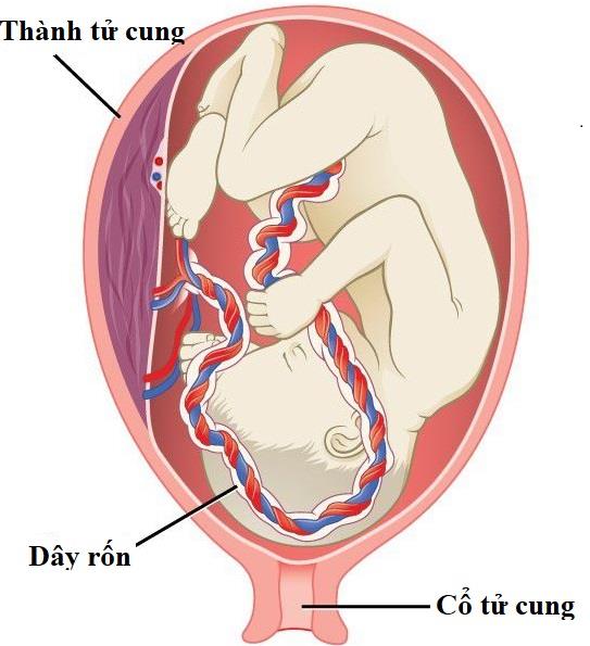
Three blood vessels from the umbilical cord are covered with Wharton's jelly
The rope is usually about 1-2 cm in diameter, and about 60 cm long. One end attaches to the center of the abdomen in the baby, the other end attaches to the center of the placenta. In some cases, the cord can attach at the edge or at the edge of the placenta. This condition causes umbilical cord disease.
After the baby is born, the cord's blood vessels close on their own. First, the arteries close first thanks to the thicker artery muscle. This advantage helps prevent blood loss from the baby to the placenta. The umbilical vein will close a little later, about 15 seconds to 3-4 minutes later. Therefore, the current trend is that a later clamping of the umbilical cord will help the baby receive more blood and iron.
The cord has no sensory nerves. So cutting the umbilical cord after birth should not hurt your baby. After the umbilical cord falls off, an umbilical cord is formed, which remains on the abdomen for life.
2. Possible unusual problems?
2.1 Prolapse of the umbilical cord:
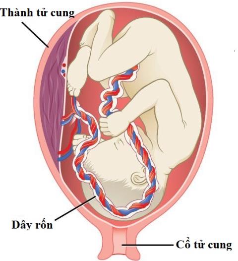
This is a complication that occurs before or during the birth of the baby. In this case, the umbilical cord prolapses through the opening of the cervix, exiting the vagina.
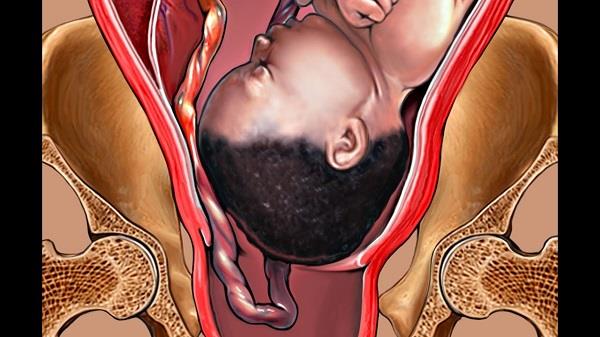
The umbilical cord prolapses through the opening of the cervix and exits the vagina
At that time, the cord can become stuck due to the pressure of the baby's body. This condition deprives the fetus of oxygen. If not treated promptly, it can even cause fetal death.
It is estimated that one out of every 300 births is an umbilical cord prolapse.
What causes umbilical cord prolapse?
- Oops broke early
- Premature birth
- Multiple pregnancy (twins, triplets, etc.)
- Too much amniotic fluid (polyhydramnios)
- The butt
- The umbilical cord is longer than usual
Doctors can diagnose cord prolapse in several ways. At birth, your doctor will use a fetal heart monitor to measure your baby's heart rate. If the cord is prolapsed, the baby may have bradycardia. Or your doctor usually checks the dilation of your cervix every 30 minutes when you're in labor. This will help detect umbilical cord prolapse in time.
Once the condition is discovered, the doctor will recommend an emergency cesarean section. This is the best approach to make sure your baby is safe.
2.2 Single artery umbilical cord (SUA):
This is a condition in which instead of the umbilical cord having two arteries, only one remains. Usually, the cord will tend to lose the left umbilical artery, about 70%.
Single-arterial umbilical cords account for about 0.4-1% of all pregnancies. This rate may increase when the mother is pregnant with twins or has diabetes.
This is a condition that can be detected with a pregnancy screening ultrasound. This condition is mostly harmless to the baby. The fetus is still nourished and developed normally. However, it has been reported that approximately 15% of fetuses with an umbilical artery will be associated with intrauterine growth retardation.
In addition, a single-arterial umbilical cord will sometimes be accompanied by a number of other chromosomal abnormalities and fetal abnormalities, such as:
- Trisomy 21 causes Down syndrome (12.8% with SUA), trisomy 18 (50% with SUA), trisomy 13 (25% with SUA)
- Birth defects such as heart, kidney, spine
- Renal aplasia: usually occurs on the side of the missing umbilical artery.
Single-arterial umbilical cords are usually detected by ultrasound. With high-resolution ultrasound, sensitivity and specificity reach 100%.
2.3 Umbilical cord wrapped around the neck:

Umbilical cord wraps can occur during pregnancy, labor, or delivery
This is the term used by medical professionals when a baby has the umbilical cord wrapped around his neck. This condition can occur during pregnancy, labor, or delivery.
The umbilical cord around the neck is a very common condition. It is estimated that one-third of babies are born completely healthy with the umbilical cord wrapped around their neck.
This condition can be detected by ultrasound. If you are diagnosed with an umbilical cord wound early in your pregnancy, it's important that you don't panic first. The cord can be removed on its own before birth. If the cord is still around the neck, the baby can actually be born safe and healthy.
Complications caused by the umbilical cord wrapping around the neck are extremely rare. The most common complication occurs when the cord is compressed (pressured) during uterine contractions. This reduces the amount of oxygenated blood reaching the baby's body and manifests itself in a decreased fetal heart rate.
With proper monitoring during delivery, the doctor can detect this problem in most cases and treat it accordingly. Therefore, most babies are born without any complications from this abnormality. If the baby's heart rate continues to drop, the doctor may recommend an emergency cesarean section to ensure the baby's safety.
In rare cases, the umbilical cord wrapped around the neck can reduce fetal movement, slow fetal growth in utero.
2.4 Knotted umbilical cord:
This condition occurs when a baby flips and rotates in the amniotic sac in the womb. Even a knotted umbilical cord can be formed even during childbirth.
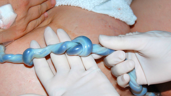
The knotted umbilical cord can be formed even during childbirth
One out of every hundred cases is a knot in the umbilical cord. This condition is more common than the umbilical cord wrapped around the baby's neck.
Risk factors for knotted umbilical cord can be due to: long umbilical cord, small fetus, twin pregnancy, etc.
The most common sign of a knotted umbilical cord is decreased fetal movement after 37 weeks. If the knot occurs during labor, the pregnancy monitor will detect an irregular heartbeat.
In fact, the condition of the umbilical cord knot is not as worrying as you think. Because the wire is protected by a layer called Wharton's jelly. This layer acts as a cushion around the blood vessel and protects the blood vessel even when it is constricted.
However, if the knot is too tight, it will interfere with the blood flow from the placenta to the baby. This condition causes the baby to be deprived of oxygen. This complication is most likely to occur during vaginal delivery. However, these cases are very rare.
There's nothing you can do to prevent the umbilical cord from getting knotted. However, you can monitor your baby's daily status through fetal movements (developing machine). In case you notice any changes in pregnancy through daily monitoring. Come to the obstetrics and gynecology medical center for direct evaluation and monitoring. If tight umbilical cord is suspected and markedly decreased fetal heart rate is detected. Your doctor may advise you to recommend an emergency cesarean section to get the baby out. Usually this is the best approach.
2.5 Umbilical cord cyst:
Umbilical cysts are sacs of fluid inside the umbilical cord. They are uncommon — less than 1 in 100 pregnancies (less than 1%) have cord cysts. This condition can be detected by ultrasound. The detection is more likely to be found in the first trimester. Most cysts found in the first trimester do not affect the baby.
If your doctor finds an umbilical cord cyst on an ultrasound, he or she may recommend additional tests such as detailed 3D, 4D ultrasound and genetic testing to check for birth defects. If the cyst is large, it may sometimes have to be removed to keep the cyst from bursting during labor.
The umbilical cord is an essential component to help the baby develop comprehensively in the womb. When there are abnormalities related to the umbilical cord, they all more or less affect the development and survival of the baby. Therefore, regular prenatal screening and check-ups are extremely important. The antenatal checkup helps to detect most abnormalities. From there, a stricter and more appropriate pregnancy management plan is proposed.
>> See more: Newborn umbilical cord: Related issues
Doctor Nguyen Trung Nghia
